Home

charter Kompleks bag cell light microscope Wings sengetøj kæmpe

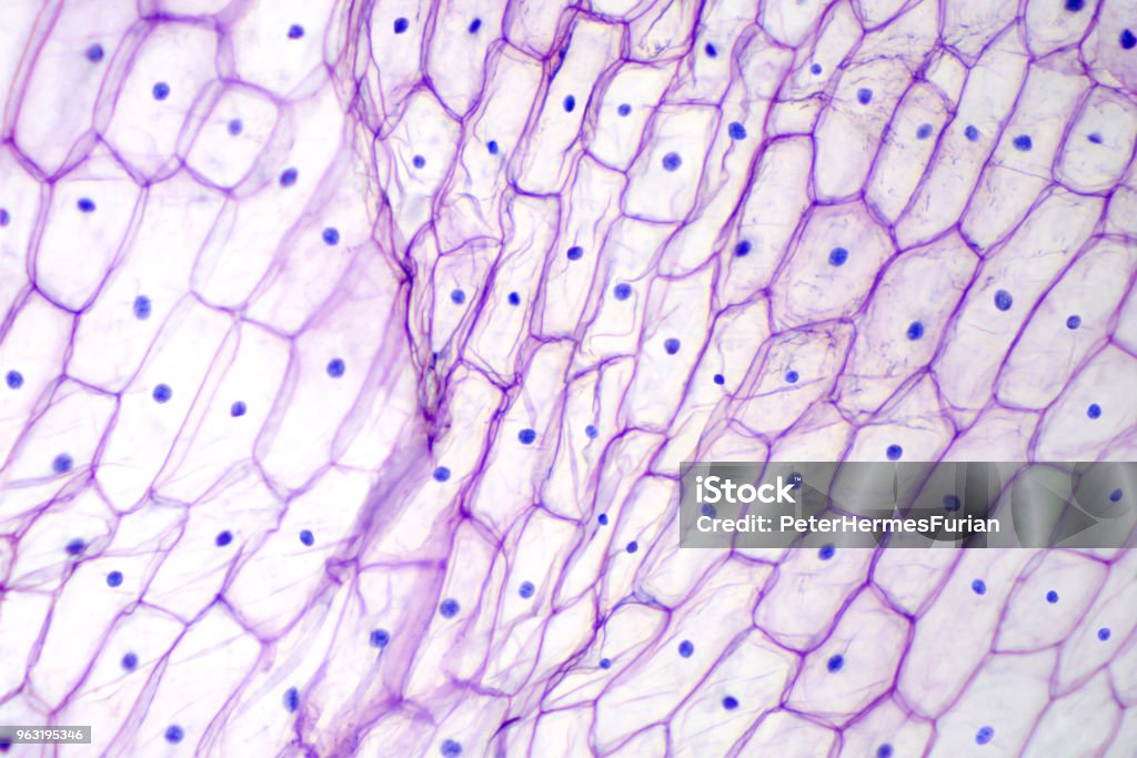
Onion Epidermis With Large Cells Under Microscope Stock Photo - Download  Image Now - Skin, Biological Cell, Peel - Plant Part - iStock
Onion Epidermis With Large Cells Under Microscope Stock Photo - Download Image Now - Skin, Biological Cell, Peel - Plant Part - iStock

Tissue Culture | Life Sciences | Solutions | Nikon Instruments Inc.

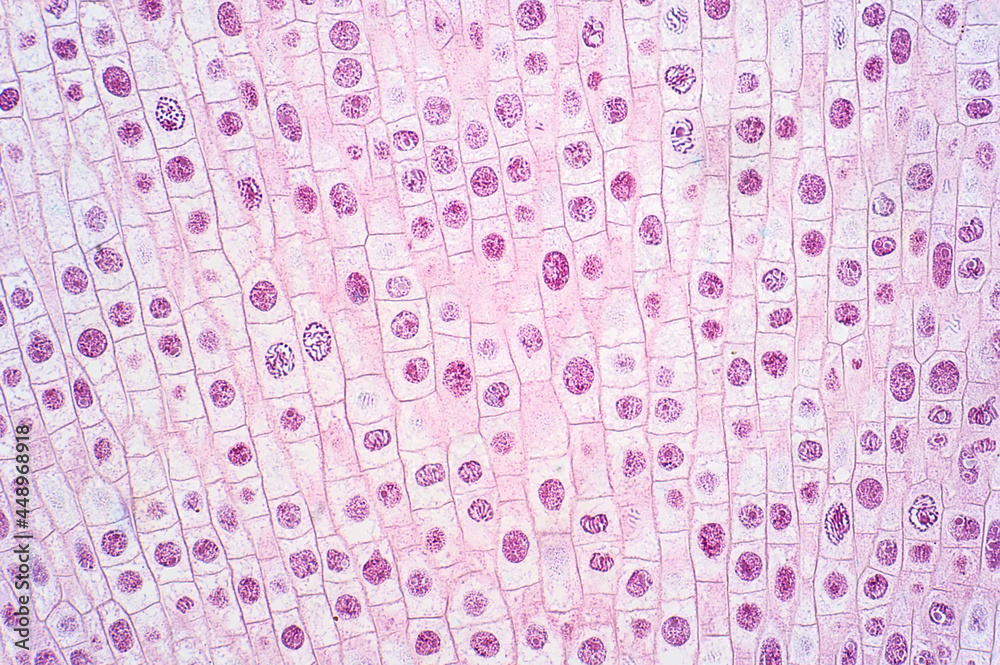
Mitosis cell of root tip of onion under the light microscope. Stock Photo |  Adobe Stock
Mitosis cell of root tip of onion under the light microscope. Stock Photo | Adobe Stock

Light microscopy | LMCB - Laboratory for Molecular Cell Biology - UCL -  London's Global University
Light microscopy | LMCB - Laboratory for Molecular Cell Biology - UCL - London's Global University

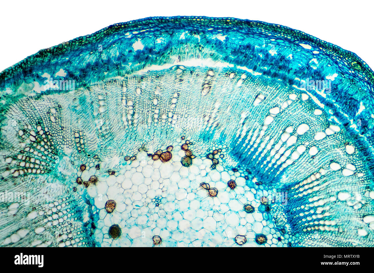
Plant cell microscope hi-res stock photography and images - Alamy

If the smallest distance a light microscope can see is 500nm, and a cell  membrane is 7nm, why can we see the cell membrane when looking through a light  microscope? - Quora
If the smallest distance a light microscope can see is 500nm, and a cell membrane is 7nm, why can we see the cell membrane when looking through a light microscope? - Quora

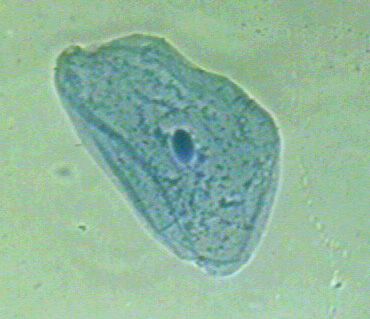
Biology 130 Lab 3 - Light Microscope Images

IntelligentDesign/RandomDrift | Plant cell, Theory of life, Photosynthesis

Cell Lab
Cell Lab

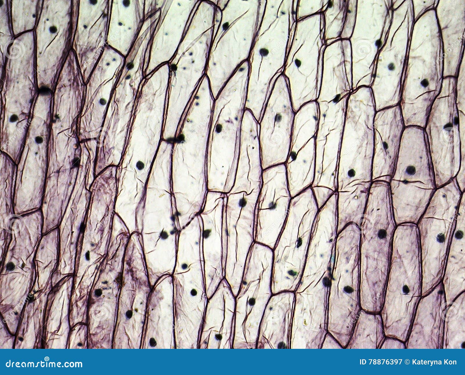
Micrograph of Onion Epidermal Cells Stock Image - Image of light, macro:  78876397
Micrograph of Onion Epidermal Cells Stock Image - Image of light, macro: 78876397

Microscopy > Light Microscopy | Campus Microscopy & Imaging Facility (CMIF)

Bright-field microscopy - Wikipedia

How to Calculate Magnification on a Light Microscope | Sciencing

Plant Cell Surface Of Leaf Under Light Microscope Stock Photo, Picture And  Royalty Free Image. Image 60142978.
Plant Cell Surface Of Leaf Under Light Microscope Stock Photo, Picture And Royalty Free Image. Image 60142978.

Innovations in Light Microscopy | Nikon's MicroscopyU

3.3 Cells under the microscope - Stile

Cell structure 1 - Animal Cells

Outline of cell biology - Wikipedia

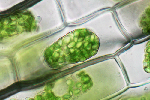
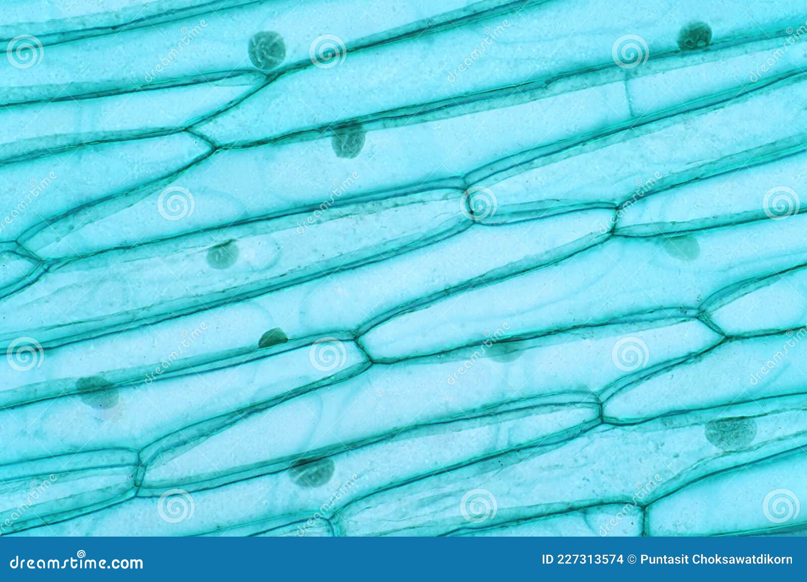
Plant Cells Under the Light Microscope View Stock Photo - Image of  chloroplast, cell: 227313574
Plant Cells Under the Light Microscope View Stock Photo - Image of chloroplast, cell: 227313574

Biology 2e, The Cell, Cell Structure, Introduction | OpenEd CUNY

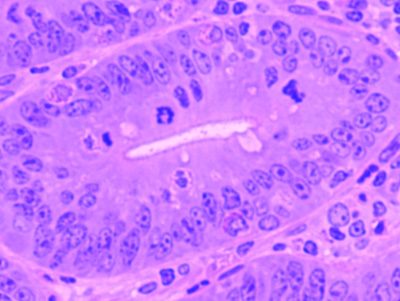
Histology Stoma Cell Under Light Microscope Stock Photo 1215942331 |  Shutterstock
Histology Stoma Cell Under Light Microscope Stock Photo 1215942331 | Shutterstock

1.5: Microscopy - Biology LibreTexts | Eukaryotic cell, Microscopy, Animal  cell
1.5: Microscopy - Biology LibreTexts | Eukaryotic cell, Microscopy, Animal cell

Learning activity 1.1: Learning about cells and microscopes

Cell Biology for the Histologist | histology

Science Plant Cells By Light Microscope Stock Photo, Picture And Royalty  Free Image. Image 110661225.
Science Plant Cells By Light Microscope Stock Photo, Picture And Royalty Free Image. Image 110661225.

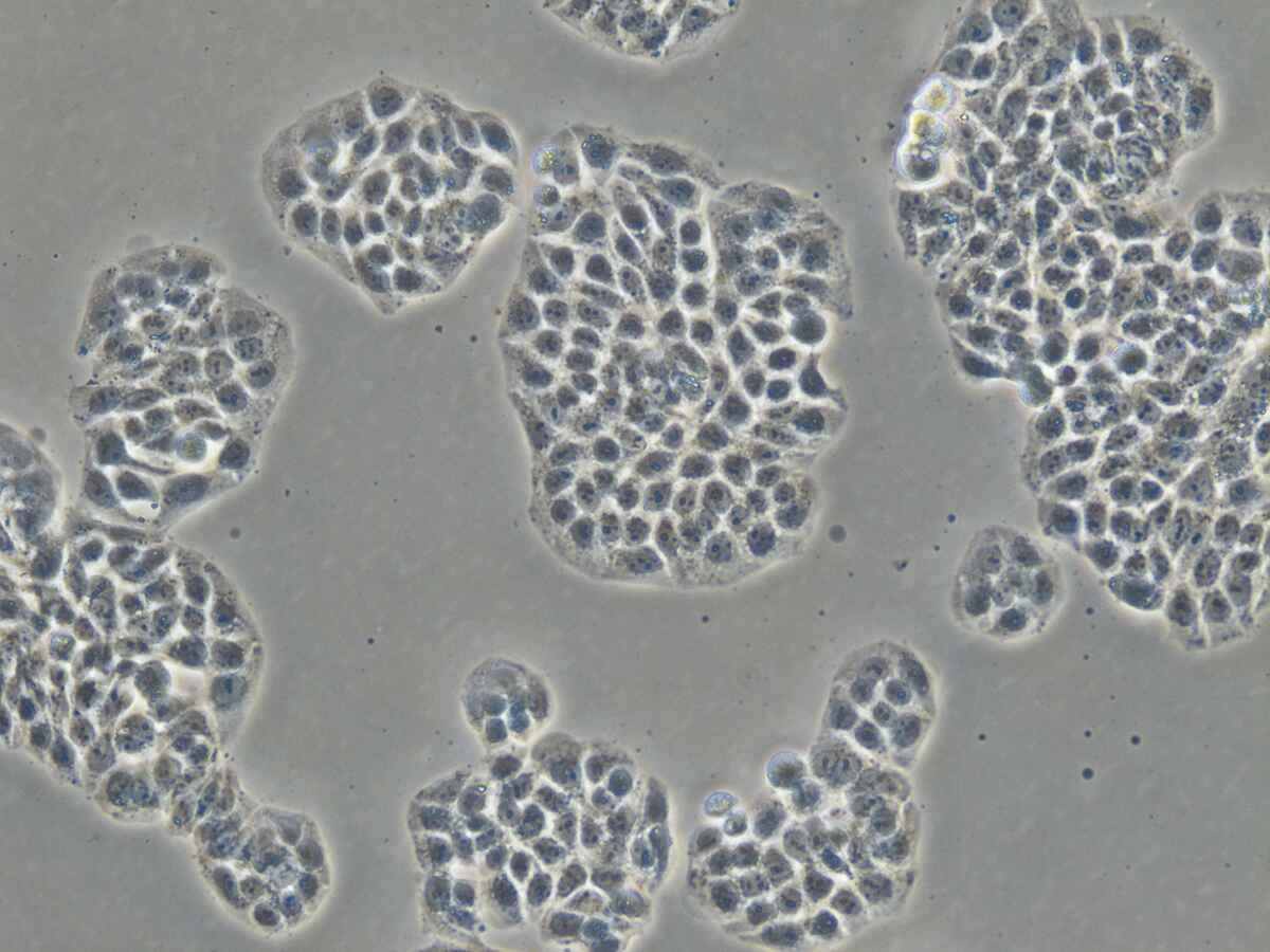
HeLa cells in culture, light microscopy - Stock Video Clip - K005/6160 -  Science Photo Library
HeLa cells in culture, light microscopy - Stock Video Clip - K005/6160 - Science Photo Library

Onion epidermis with large cells under light microscope Greeting Card by  Peter Hermes Furian
Onion epidermis with large cells under light microscope Greeting Card by Peter Hermes Furian

Onion epidermis with large cells under light microscope Zip Pouch by Peter  Hermes Furian - Fine Art America
Onion epidermis with large cells under light microscope Zip Pouch by Peter Hermes Furian - Fine Art America

Microscopy
Microscopy